



Systomus rubripinnis (Valenciennes, 1842), commonly known as the Javaen barb or Red Cheek Barb, is a tropical freshwater cyprinid belonging to the family Cyprinidae within the order Cypriniformes. In the Malay language this species is known as Ikan Pipi Merah (Ng et al., 2017). The species is benthopelagic and inhabits freshwater environments, with reported preferences for a pH of approximately 6.0–6.5 and temperatures of 22–25°C (Riehl and Baensch, 1991). Its distribution extends across tropical Asia, particularly the Malay Peninsula and Indonesia, as well as the Mekong, Mae Khlong, and Chao Phraya river basins. In Malaysia, S. rubripinnis is consumed as a food fish, although its availability and market value fluctuate seasonally. It’s comparatively high protein content relative to silver barb and tilapia further supports its nutritional and commercial relevance (Hadisusanto and Suryaningsih, 2011). The species can be distinguished by characteristic red pigmentation of the eye iris and operculum and brownish coloration at the distal portions of the fins. Despite its relatively broad occurrence in Southeast Asia, S. rubripinnis has reportedly declined in some areas, with local disappearance of subpopulations in Java. However, insufficient population data have limited its formal assessment as an endangered species, emphasizing the importance of improving knowledge of its reproductive biology and developing approaches for controlled propagation.
The ecology of S. rubripinnis is closely associated with flowing freshwater habitats. The species occurs in rivers of different sizes but is particularly common in smaller streams, canals, and floodplains, while its occurrence in impoundments is generally associated with connected flowing waters (Harvolk et al., 2014). Reproductive activity is linked to seasonal hydrological conditions, with adults moving toward seasonally flooded habitats during the onset of the rainy season. Young-of-the-year individuals have been observed in streams during July and August, whereas adults leave floodplain habitats as floodwaters recede during December or January (Horn et al., 2011). In Thailand, S. rubripinnis is also harvested from the wild for the ornamental fish trade (Sassa-Deepaeng et al., 2024). Such dependence on seasonal environmental conditions and continued extraction from natural populations may constrain the consistent availability of the species for both food and ornamental purposes, highlighting the need for reliable captive breeding and seed production.
Previous investigations have provided useful information on the reproductive ecology of Systomus and related cyprinid fishes. Reproductive characteristics of Systomus species were examined in the Serayu River, while the reproductive biology of Osteochilus hasselti and P. orphoides was investigated in the Pelus River, Banyumas (Susatyo et al., 2022; Iswantari et al., 2021). However, these studies primarily addressed natural reproduction under native environmental conditions and therefore provide limited guidance for controlled hatchery production. Hormone-induced artificial breeding was successfully applied to Javaen barbs, indicating the potential of reproductive biotechnology for overcoming some limitations of natural spawning (Yu Abit et al., 2021). Nevertheless, reproductive readiness and gonadal maturation remain strongly influenced by seasonal conditions, which can result in inconsistent spawning and inadequate nursery production. A further critical limitation is the lack of a detailed chronological and morphological description of embryonic and early larval development in S. rubripinnis. Without reliable information on the timing of cleavage, blastula formation, gastrulation, segmentation, hatching, and yolk-sac absorption, hatchery operators have limited biological benchmarks for egg monitoring, hatch management, and determining the appropriate onset of external feeding.
Accordingly, the central problem addressed by this study is the insufficiently characterized early developmental biology of S. rubripinnis and the resulting limitation in establishing predictable captive propagation protocols. In particular, it remains unclear whether hormone-induced spawning can provide viable offspring under captive conditions and how rapidly the fertilized eggs progress through successive embryonic stages before hatching and early larval development. The study therefore asks whether S. rubripinnis can be successfully induced to spawn using a standardized hormone protocol and what are the chronological and morphological characteristics of its embryonic and early larval development under hatchery conditions. It was hypothesized that hormone-induced spawning would produce viable fertilized eggs and that embryonic development would proceed through distinct, temporally identifiable morphological stages culminating in successful hatching and subsequent depletion of the yolk reserve.
The objective of this study was to induce spawning in sexually mature S. rubripinnis under captive conditions and characterize the chronological progression and morphological features of its embryonic and early larval development from fertilization to the initial feeding stage. Particular emphasis was placed on identifying the timing of major developmental transitions and the duration of yolk-sac utilization. The novelty of the study lies in integrating successful hormone-induced breeding with systematic microscopic documentation of the early developmental sequence of S. rubripinnis, thereby establishing species-specific developmental benchmarks that are currently limited in the literature. By linking spawning induction with the timing of embryonic differentiation, hatching, larval pigmentation, and yolk-sac absorption, this study provides a practical developmental framework for hatchery management. The findings are expected to contribute to more predictable seed production, improve the timing of first exogenous feeding and larval husbandry, and support the broader domestication and aquaculture development of S. rubripinnis while potentially reducing dependence on seasonally available wild stocks.
2. Materials and methods
2.1 Ethical approval
No ethical approval was required to conduct the study.
2.2 Study site and experimental period
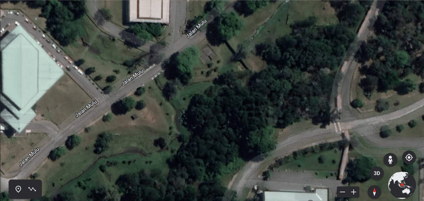
Broodstock of S. rubripinnis were collected from a natural stream located within the Universiti Putra Malaysia Sarawak (UPMS), Malaysia (3.206700°N, 113.091344°E; Figure 1). The stream had a maximum width of approximately 5 m and a maximum depth of 2 m. Fish were captured using gill nets and scoop nets. Non-target species were immediately released at the collection site, whereas S. rubripinnis individuals were retained for broodstock selection. Sexually mature individuals were identified by external reproductive characteristics, including the presence of milt upon gentle abdominal pressure in males and reddish gonopores in females. Selected individuals were transported to the Aquatic Biotech Laboratory and Wet Laboratory, Academic Centre Complex, Department of Animal Science and Fisheries, Faculty of Agricultural Science and Forestry, Universiti Putra Malaysia Sarawak. Induced-breeding trials were conducted from 14 to 16 October 2022.
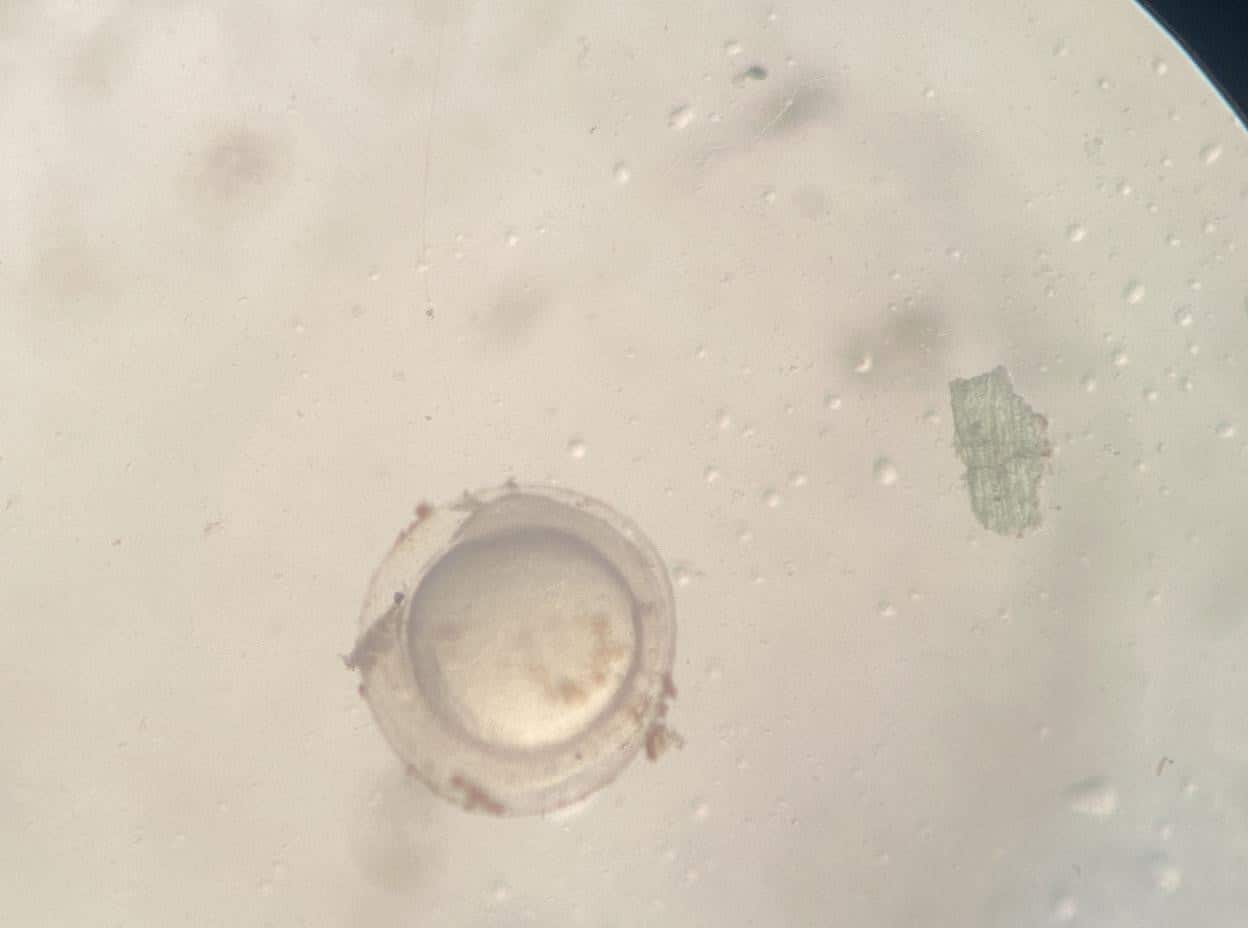
Figure 1. Geographic location of the Systomus rubripinnis broodstock sampling site at Universiti Putra Malaysia Sarawak, Malaysia.
2.3 Water quality management
Dechlorinated tap water was used throughout broodstock maintenance and the breeding experiments. An anti-chlorine treatment was added whenever water was replaced to eliminate residual chlorine. Water-quality conditions were maintained within the ranges considered suitable for S. rubripinnis broodstock and early developmental stages (Table 1). Dissolved oxygen, ammonia, carbon dioxide, salinity, and pH were monitored and maintained at the specified levels throughout the experimental period.
Table 1. Target water-quality parameters maintained during broodstock conditioning and induced breeding of Systomus rubripinnis.
| Parameter | Values |
| Salinity | 0 ppt |
| Dissolved oxygen | Below 6 mg/litre |
| Ammonia | Below 0.5 mg/litre |
| Carbon dioxide | Below 10 mg/litre |
| pH level | 6.8-7.8 |
2.4 Broodstock preparation and hatchery management
Sexually mature male and female broodstock were separated into two aerated 120-L plastic tanks according to sex. The fish were fed commercial floating starter pellets twice daily and acclimatized for one week before hormone induction. Prior to hormone administration, broodstock were anesthetized using tricaine methanesulfonate (MS-222) at 65 mg L⁻¹ to minimize handling stress and excessive movement during injection. Induced spawning was performed using Ovaprim® (Syndel), a salmon gonadotropin-releasing hormone analogue (sGnRHa) preparation containing 20 µg GnRH and 10 mg domperidone per mL. The use of Ovaprim for stimulating spawning in cyprinid fishes has previously been demonstrated (Abit et al., 2024; Ashraf et al., 2024; Ali et al., 2015). The materials required for the induced-breeding procedure included a 0.5-mL syringe, a fine injection needle, Ovaprim® (Syndel), and a weighing scale.
2.4.1 Hormone administration and induced spawning
Each female broodstock was weighed individually before hormone administration. The required hormone volume was calculated according to the following equation:
Hormone volume (mL) = body weight (g) / 1000 × 0.5 mL kg⁻¹
The calculated dose was drawn into a 0.5-mL syringe and administered by intramuscular injection near the anterior region of the dorsal fin. Following injection, the female was transferred to the breeding tank. Male broodstock received half the hormone dose administered to the female (0.25 mL kg⁻¹) and were subsequently transferred to the same breeding tank. Spawning occurred approximately 12–24 h after hormone administration. The fertilized eggs were subsequently maintained under controlled hatchery conditions for embryonic-development observations.
2.5 Documentation and analysis of embryonic and larval development
Embryonic and larval development was monitored from fertilization through hatching and the subsequent early larval period. Fertilized eggs and larvae were examined using a Leica CME dissecting microscope equipped for approximately 40–100× magnification. Sequential photographs were taken to document morphological changes throughout development. During the earliest developmental period, observations were conducted at 15-min intervals, whereas observations were subsequently made at approximately 1-h intervals until hatching. Developmental stages were identified according to characteristic morphological events, including cleavage, blastula formation, gastrulation, segmentation, tail-bud development, organogenesis, and hatching. Post-hatching larvae were also observed to document changes in pigmentation, eye development, and yolk-sac utilization. The duration of each developmental stage was determined from the recorded time elapsed after fertilization.
3. Results
3.1 Early embryonic and larval development of S. rubripinnis
The embryonic development of S. rubripinnis proceeded through six major developmental periods: zygote, cleavage, blastula, gastrula, segmentation, and hatching. The transition between stages was identified from distinct morphological changes observed microscopically. During the first three developmental periods, observations were conducted at 15-min intervals to capture the rapid early cleavage events, whereas subsequent observations were performed at approximately 1-h intervals until hatching. The complete sequence from fertilization to hatching was completed in approximately 14 h (Table 2).
Table 2. Chronology and major morphological characteristics of early embryonic development in Systomus rubripinnis.
| Developmental period | Approximate time post-fertilization | Major developmental characteristics |
|
Zygote |
0–0.75 h |
Fertilized egg remains at the zygote stage; the chorion expands and separates from the yolk, while cytoplasmic streaming establishes the blastodisc at the animal pole. |
|
Cleavage |
0.75–2 h |
The first cleavage occurs approximately 45 min after fertilization, followed by rapid and initially synchronous cell divisions within the blastodisc. |
|
Blastula |
2–6 h |
The blastodisc develops into a multicellular blastula; the yolk syncytial layer forms, the mid-blastula transition occurs, and epiboly begins. |
|
Gastrula |
6–9 h |
Epiboly progresses, accompanied by involution, convergence, and extension, resulting in formation of the primary germ layers and embryonic axis. |
|
Segmentation |
9–14 h |
Somites develop sequentially, the embryo elongates, the tail bud becomes prominent, and early organogenesis occurs. |
|
Hatchling |
~14 h |
Embryonic motility increases, the chorion ruptures, and actively moving hatchlings emerge from the egg envelope. |
3.1.1 Zygote period
The zygote period extended from fertilization until the onset of the first cleavage, approximately 45 min post-fertilization. The fertilized eggs were spherical, demersal, adhesive, translucent to mildly brownish, and had an approximate diameter of 0.75 mm. Within 7–10 min after fertilization, the chorion expanded and separated from the newly fertilized egg, becoming distinctly elevated around the yolk. Concurrent cytoplasmic streaming toward the animal pole resulted in the differentiation of the clearer, yolk-rich vegetal cytoplasm from the non-yolky cytoplasmic blastodisc. This spatial organization persisted during the initial stages of cleavage (Figure 2).
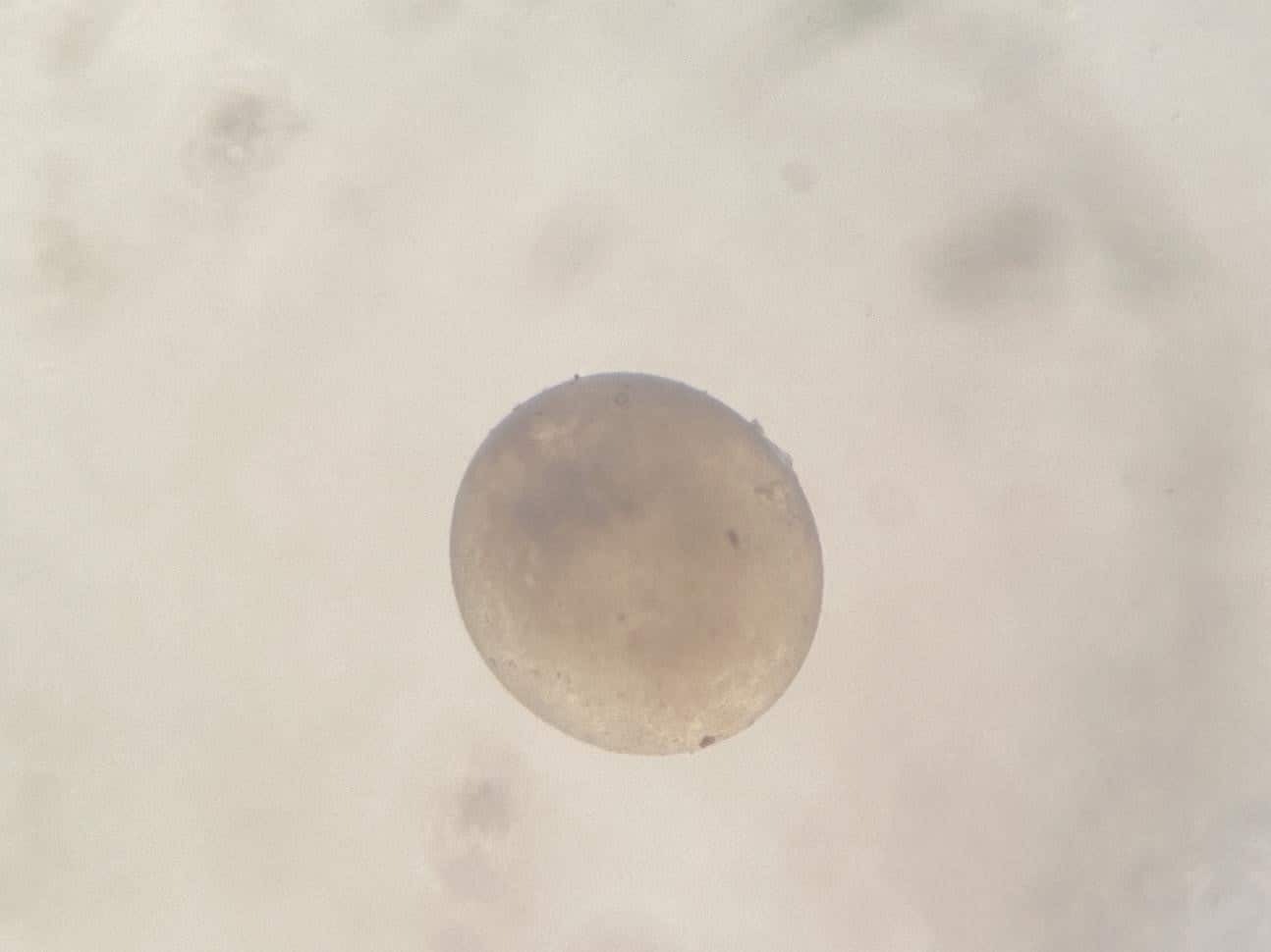
Figure 2. Representative microscopic features of the zygote period of Systomus rubripinnis from fertilization to the onset of first cleavage.
3.1.2 Cleavage period
The first cleavage occurred approximately 45 min after fertilization. The cleavage furrow originated near the animal pole and progressed rapidly toward the vegetal pole, dividing only the blastodisc while leaving the yolk undivided. The first vertical cleavage produced two blastomeres of approximately equal size. Subsequent cleavage divisions occurred rapidly, with cell cycles progressing through successive stages and initially displaying a high degree of synchrony. By approximately 2 h post-fertilization, the blastodisc had undergone extensive cellular division and transitioned toward the blastula stage (Figure 3).
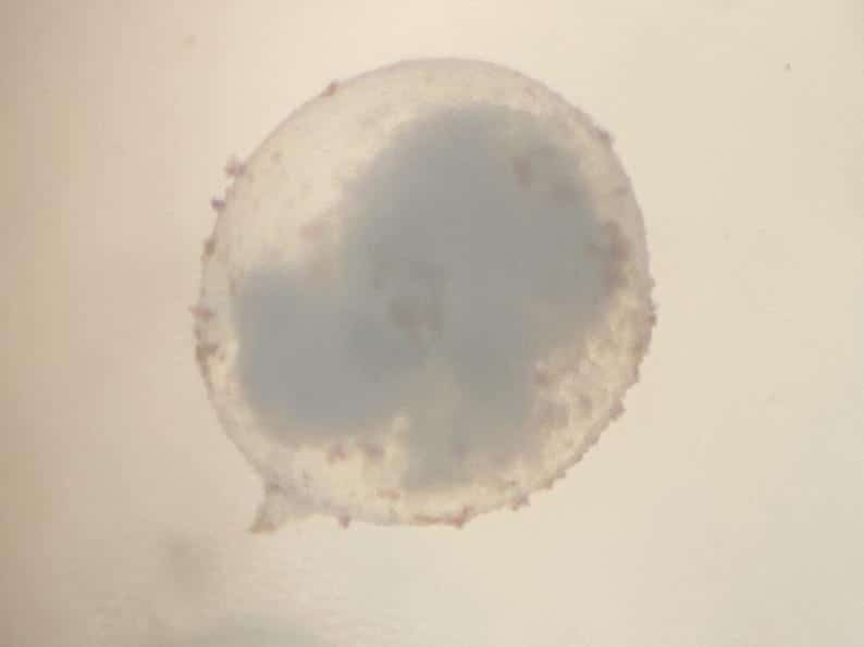
Figure 3. Representative microscopic features of the cleavage period of Systomus rubripinnis from the first cleavage to approximately 2 h post-fertilization.
3.1.3 Blastula period
The blastula period occurred approximately between 2 and 6 h post-fertilization. At the beginning of this period, the blastodisc had developed into a compact multicellular structure, corresponding to the advanced cleavage stages. The major developmental events included the formation of the yolk syncytial layer, the mid-blastula transition, and the initiation and progression of epiboly. During this period, cell division progressively shifted from synchronous to asynchronous activity. Epiboly subsequently continued into the gastrula period as the blastoderm expanded over the yolk cell (Figure 4).
Figure 4. Representative microscopic features of the blastula period of Systomus rubripinnis between approximately 2 and 6 h post-fertilization.
3.1.4 Gastrula period
Gastrulation occurred approximately between 6 and 9 h post-fertilization. Epiboly continued throughout this period, accompanied by coordinated morphogenetic cell movements, including involution, convergence, and extension. These processes resulted in progressive organization of the primary germ layers and establishment of the embryonic axis. The embryo became increasingly differentiated and structurally organized as development progressed toward segmentation (Figure 5).
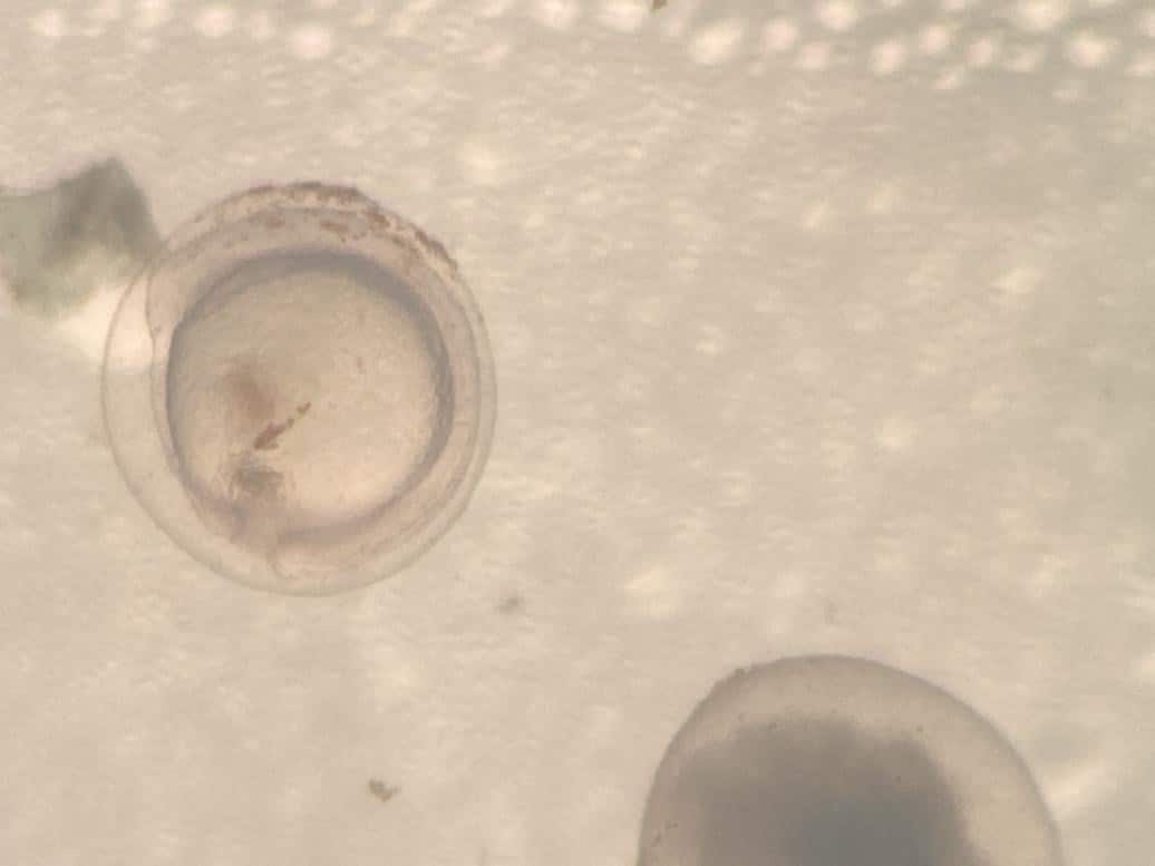
Figure 5. Representative microscopic features of the gastrula period of Systomus rubripinnis between approximately 6 and 9 h post-fertilization.
3.1.5 Segmentation period
The segmentation period occurred approximately between 9 and 14 h post-fertilization. Following completion of epiboly, the first somatic furrows became apparent, marking the initiation of somite formation, while the tail bud became increasingly distinct. Somites developed sequentially in an anterior-to-posterior direction, and the embryo progressively elongated. Concurrently, the tail bud became more prominent and rudiments of major organs began to differentiate. Several distinct somite stages were documented during this period (Figure 6). At the 12-somite stage, somites were clearly identifiable along the developing embryo. At approximately 16 somites, the somites began to exhibit a chevron-like arrangement, while the yolk extension became more apparent as the yolk cell acquired a kidney- or bean-like configuration. The Kupffer’s vesicle was also visible, and the tail bud became increasingly prominent. Subsequent observations documented the 18-, 20-, and 30-somite stages, demonstrating progressive segmentation and embryonic elongation.
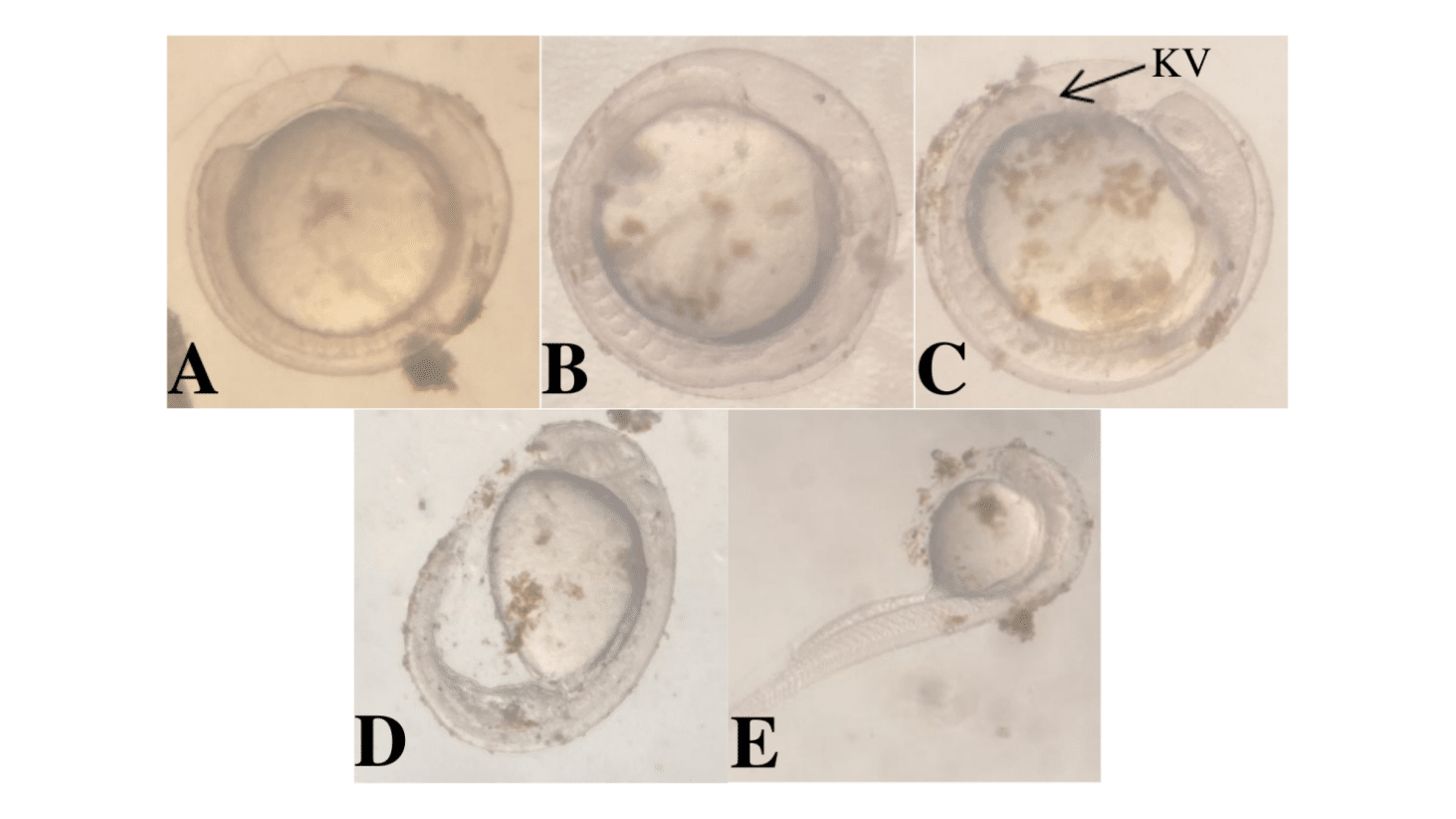
Figure 6. Representative developmental stages during segmentation of Systomus rubripinnis. (A) 12-somite stage; (B) 16-somite stage showing chevron-shaped somites, kidney- or bean-shaped yolk extension, Kupffer’s vesicle, and a prominent tail bud (arrow); (C) 18-somite stage; (D) 20-somite stage; and (E) 30-somite stage.
3.1.6 Hatching and early larval development
Hatching commenced at approximately 14 h post-fertilization. At this stage, the embryo exhibited pronounced movement and progressive separation of the developing tail from the yolk mass. The embryos occasionally twisted within the chorion, and the pectoral-fin buds were relatively elongated. Cardiac activity was clearly visible, with vigorous heartbeats and apparent blood circulation. Increasing embryonic movement ultimately contributed to rupture of the chorion and emergence of the hatchlings.
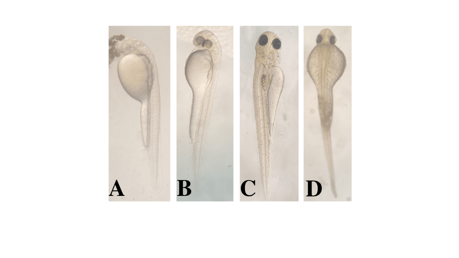
Figure 7. Early larval development of Systomus rubripinnis following hatching. (A) Newly hatched larva; (B) 12-h-old larva showing increasing eye pigmentation; (C) 24-h-old larva showing the onset of dorsal pigmentation and clearly differentiated eyes; and (D) 24-h-old larva in dorsal view.
Following hatching, the larvae remained associated with their yolk reserves and underwent progressive morphological differentiation. Newly hatched larvae exhibited limited pigmentation, whereas eye pigmentation became increasingly evident by 12 h post-hatching. At 24 h post-hatching, pigmentation was visible dorsally and the eyes were clearly differentiated (Figure 7). These observations demonstrate progressive morphological development during the initial larval period following hatching.
4. Discussion
Successful captive propagation of freshwater cyprinids depends on the ability to synchronize spawning with appropriate broodstock conditioning and to maintain suitable conditions during embryonic and larval development. Previous work has demonstrated the feasibility of induced breeding in Javaen barb, providing an important basis for developing controlled reproductive protocols for S. rubripinnis (Iswantari et al., 2021). The present study extends this knowledge by combining hormone-induced spawning with detailed observation of the subsequent embryonic sequence, thereby providing a chronological framework for early development from fertilization to hatching and early larval differentiation.
4.1 Induced breeding of S. rubripinnis
The present study demonstrated that induced spawning of sexually mature S. rubripinnis could be achieved using Ovaprim® (Syndel), containing salmon gonadotropin-releasing hormone analogue (sGnRHa) and domperidone. A dose of 0.5 mL kg⁻¹ was administered to females, while males received half of the female dose (0.25 mL kg⁻¹). The female dose falls within the range of 0.4–0.5 mL kg⁻¹ body weight recommended for female cyprinid broodstock by Ashraf et al. (2024), while the present results indicate that a half-dose of 0.25 mL kg⁻¹ was sufficient for male S. rubripinnis. This difference between sexes is consistent with the common use of reduced hormone doses in males because spermiation generally requires less endocrine stimulation than final oocyte maturation and ovulation.
The successful response at the applied dose also indicates that hormone-induced spawning may provide a practical means of overcoming some of the seasonal limitations associated with natural reproduction in S. rubripinnis (Zamri et al., 2022). This is particularly relevant because reproductive activity in cyprinids is strongly influenced by environmental and seasonal conditions. By inducing spawning under controlled conditions, hatchery production can potentially be made more predictable, provided that broodstock have reached an appropriate stage of gonadal maturity (Siddique et al., 2022; Migaud et al., 2013). Broodstock handling and hormone administration are important components of successful induced breeding. In the present experiment, fish were anesthetized with MS-222 at 65 mg L⁻¹ before injection to reduce excessive movement and handling-related stress (Park, 2019).
Intramuscular administration using a 0.5-mL syringe allowed the calculated hormone dose to be delivered accurately while minimizing unnecessary manipulation. Appropriate needle size and careful injection technique are particularly important because mechanical injury can increase physiological stress and compromise broodstock condition. Methylene blue was also used in the breeding system to reduce the risk of fungal and bacterial contamination associated with eggs and minor injection-related injuries. In addition, removal of unfertilized or deteriorating eggs helped maintain water quality during embryonic development (Li et al., 2023).
4.2 Embryonic and early larval development of S. rubripinnis
The present study identified six principal developmental periods in S. rubripinnis: zygote, cleavage, blastula, gastrula, segmentation, and hatching/early larval development. This developmental classification is broadly comparable with the sequence reported for rosy barb, Puntius conchonius, by Bhattacharya et al. (2005), although differences in the number and definition of developmental stages have been reported among cyprinid species. For example, Basak et al. (2014) described eight developmental stages in silver barb, Barbodes gonionotus. Such variation may reflect differences in species-specific developmental rates, environmental conditions, observational resolution, and the criteria used to delineate developmental stages.
Fertilized S. rubripinnis eggs were spherical, demersal, adhesive, translucent to mildly brownish, and approximately 0.75 mm in diameter. The chorion became distinctly elevated within 7–10 min after fertilization, followed by cytoplasmic redistribution and formation of the blastodisc at the animal pole. The first cleavage occurred approximately 45 min after fertilization, indicating a relatively rapid onset of embryogenesis. Early cleavage divisions progressed rapidly within the blastodisc, while the yolk remained undivided. By approximately 2 h post-fertilization, development had progressed to the blastula period, during which the yolk syncytial layer formed and epiboly commenced (Ghosh et al., 2022).
The transition from blastula to gastrula occurred at approximately 6 h post-fertilization. During gastrulation, continued epiboly was accompanied by involution, convergence, and extension, resulting in the organization of the primary germ layers and establishment of the embryonic axis. These developmental processes represent a major transition from relatively rapid cellular proliferation to coordinated tissue organization. The subsequent segmentation period, beginning at approximately 9 h post-fertilization, was characterized by progressive embryonic elongation, sequential somite formation, tail-bud development, and early organogenesis. The observation of 12-, 16-, 18-, 20-, and 30-somite stages demonstrates the rapid structural differentiation occurring within only a few hours before hatching (Webb and Miller, 2006).
Hatching began at approximately 14 h post-fertilization, indicating that S. rubripinnis completes its major embryonic development within a relatively short period under the experimental conditions. Increasing embryonic movement, visible cardiac activity, tail development, and progressive separation of the tail from the yolk mass preceded rupture of the chorion. The rapid embryonic development observed here is particularly important for hatchery management because developmental events occur within narrow temporal windows, making frequent monitoring necessary during the period immediately preceding hatching (Zadmajid et al., 2018).
Following hatching, morphological differentiation continued rapidly. Eye pigmentation increased during the first 12 h after hatching, while dorsal pigmentation became evident and the eyes were clearly differentiated by 24 h. The yolk reserve was reported to be largely depleted by approximately 72 h post-hatching. Thus, the first three days after hatching represent a critical transition period in which endogenous yolk reserves progressively decline and larvae become increasingly dependent on external food resources. The 72-h yolk-utilization period therefore provides a practical biological benchmark for hatchery operators to plan the initiation and intensification of exogenous feeding (Yanes-Roca et al., 2012).
5. Conclusions
This study demonstrated the successful hormone-induced breeding and captive embryonic development of S. rubripinnis. Fertilized eggs were spherical, demersal, adhesive, translucent to mildly brownish, and approximately 0.75 mm in diameter. Embryonic development progressed through six major stages—zygote, cleavage, blastula, gastrula, segmentation, and hatching—with hatching commencing approximately 14 h after fertilization. Larvae subsequently developed progressively during the first 72 h, corresponding to substantial yolk-sac utilization and the transition toward external feeding. These species-specific developmental benchmarks provide useful guidance for egg monitoring, hatching management, and timing of first feeding, thereby supporting the development of reliable hatchery and aquaculture protocols for S. rubripinnis.
