


Microalgae are photosynthetic microorganisms that produce high-value biomass due to their simple cellular structure, efficient nutrient uptake, and adaptability to diverse aquatic environments (Sarıtaş et al., 2025; Natrah et al., 2011). They synthesize essential biomolecules, including lipids, proteins, carbohydrates, pigments, and vitamins, making microalgae a valuable resource for industries such as cosmetics, pharmaceuticals, bioenergy, and aquaculture (Ahmad et al., 2024; Fernandes et al., 2021; Becker, 2007). In shrimp aquaculture, microalgae are widely used as live food for mollusks, crustaceans, and fish larvae because of their high nutritional value and digestibility (Michels, 2015). Species such as Tetraselmis suecica, Isochrysis galbana, and Skeletonema costatum are commonly utilized in larviculture, serving not only as a dietary source but also as biological agents that enhance water quality (Ma and Hu, 2024). Microalgae improve culture conditions by increasing dissolved oxygen through photosynthesis, assimilating nitrogenous waste, and suppressing pathogens via competition and quorum sensing inhibition (QSI) (Baharuddin et al., 2024; Han et al., 2019). Certain species can disrupt bacterial quorum sensing systems, providing a natural biocontrol mechanism that reduces virulence and biofilm formation, thus offering an alternative to antibiotic use in aquaculture (Jiang et al., 2019).
Shrimps, particularly penaeid species like Penaeus monodon, are economically significant in aquaculture and have a complex developmental lifecycle that includes nauplius, zoea, mysis, and postlarval stages (Vance and Rothlisberg, 2020). During the zoea and mysis phases, shrimp rely heavily on microalgae and phytoplankton for nutrition (Brown et al., 1997). Their anatomical adaptations, including a cephalothorax and abdomen with specialized feeding and locomotion appendages, enable them to thrive in various aquaculture environments (Ruppert et al., 2004). However, intensive culture practices in tropical conditions (25 °C to 32 °C), common in Malaysian shrimp farms, have increased the risk of disease outbreaks (Nazarudin et al., 2025; Brumfield et al., 2023; Pragthong et al., 2020).
Despite the widespread use of imported strains, research on locally sourced microalgae remains limited, even though such strains may be better adapted to native environments. As autotrophic organisms, microalgae thrive in diverse marine and freshwater habitats, providing both nutritional and bioremediation benefits to aquaculture systems. To address this knowledge gap, this study aimed to isolate, identify, and characterize indigenous microalgae from a Malaysian shrimp farm using both morphological and molecular techniques. Among the isolates, Picochlorum sp. and Synechococcus elongatus were selected for further evaluation based on their distinct physiological traits and ecological relevance. Picochlorum sp. is a robust green microalga known for its high salinity tolerance, rapid growth, and production of bioactive compounds, making it well-suited to the variable conditions of shrimp ponds (Krishnan et al., 2025; Foflonker et al., 2018). Meanwhile, S. elongatus, a fast-growing cyanobacterium, has been extensively studied for its high photosynthetic efficiency and simple genetic architecture, making it an excellent model for biotechnological applications and nutrient recycling in aquaculture (Meng et al., 2025; Mills et al., 2022). Together, these traits align with the goals of sustainable aquaculture, particularly in enhancing shrimp health, improving water quality, and reducing reliance on synthetic additives.
The study explores the diversity and functional potential of indigenous microalgae from Malaysian shrimp ponds, focusing on their adaptability and suitability for tropical aquaculture systems. It is anticipated that indigenous species will exhibit distinct morphological and genetic characteristics compared to widely used commercial strains. Selected isolates, such as Picochlorum sp. and S. elongatus, are expected to demonstrate high growth performance, nutrient uptake efficiency, and quorum-sensing inhibition capabilities. By addressing these hypotheses, the research aims to provide a scientific basis for utilizing native microalgae as functional components in sustainable shrimp farming. The outcomes of this study are expected to have significant implications for the aquaculture industry, as insights gained from identifying and evaluating locally adapted microalgae will support the development of region-specific biological resources for shrimp hatcheries and grow-out systems. These findings will contribute to reducing dependence on imported strains, improving pond ecology, and promoting environmentally responsible aquaculture practices aligned with antimicrobial resistance (AMR) mitigation goals in tropical production systems.
2. Materials and Methods
2.1 Ethics declarations
Not applicable.
2.2 Sample collection and culture media preparation
This study was conducted from May, 2022, to December 1, 2022, at Zaiyadal Aquaculture Sdn. Bhd., located in the northwestern region of Selangor, Malaysia (3.680000°N, 100.970000°E) (Figure 1). Water samples were collected from five shrimp culture sites within the facility, comprising both wastewater (WW) and shrimp pond water (PW) sources. The specific sampling points were: WW 1 (3.678077°N, 100.965825°E), WW 1.2 (3.677671°N, 100.966266°E), PW 1 (3.677629°N, 100.965872°E), PW 2 (3.677509°N, 100.965326°E), and PW 3 (3.677303°N, 100.965172°E) (Figure 2). Samples were obtained using a plankton net (10-30 cm) with a 60 µm mesh size. The collected water samples were transferred into clean bottles with loose-fitting caps to allow for air exchange and were transported at room temperature for immediate laboratory processing. For cultivation, Conway medium, enriched with and without silicate, was prepared according to Walne (1970) using filtered seawater supplemented with nutrients, trace metals, and vitamins to support diverse microalgae types, including diatoms.
2.3 Microalgae isolation and purification
Microalgae were isolated using the single-cell micropipette technique under aseptic conditions, following previously described procedures (Andersen, 2005; Guillard, 2005; Stein, 1980). Individual microalgal cells were separated from pond water samples with a modified Pasteur pipette under a light microscope and transferred into sterilized Conway medium. This isolation process was repeated several times to ensure single-cell purity before transferring each cell into 10 mL culture tubes. The cultures were incubated under a 12:12 h light–dark cycle at 25 °C, illuminated by fluorescent light at 60–75 µmol photons m⁻² s⁻¹.
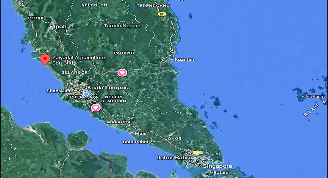
Figure 1. Zaiyadal Aquaculture Sdn. Bhd located in West Malaysia.
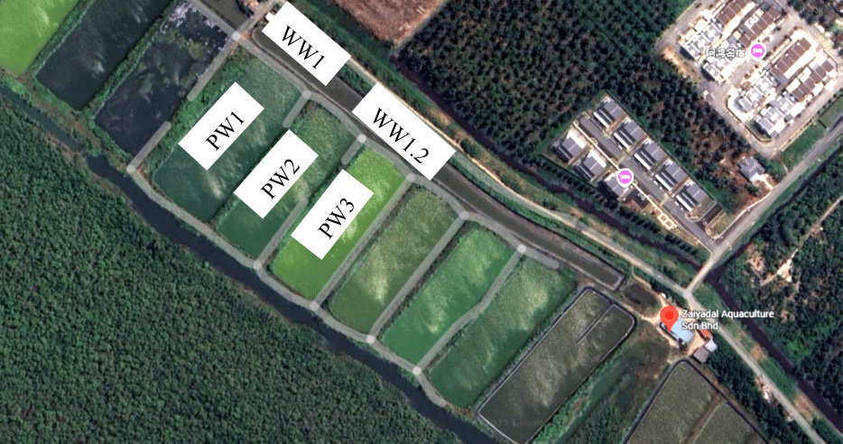
Figure 2. Sampling locations at Zaiyadal Aquaculture Sdn. Bhd., Selangor, Malaysia. Water samples were collected from three water pond sites (PW1, PW2, PW3) and two wastewater discharge channels (WW1, WW1.2). WW = Wastewater; PW = Pond water.
Purification of the isolates was achieved through repeated streaking on Conway agar plates (1.5% agar) and subsequent incubation under similar conditions, as adapted from Andersen (2005) and Torzillo and Vonshak (2013). Colonies that appeared after approximately three weeks were sub-cultured multiple times to eliminate bacterial contaminants. Antibiotics (streptomycin and penicillin) were added during the early purification stages to suppress bacterial growth (Day et al., 2012; Kooistra et al., 2008). Once axenic status was confirmed microscopically, the isolates were transferred into liquid Conway medium and gradually upscaled for further analyses.
All purified cultures were maintained at 30 ppt salinity, 25 ± 2 °C, and a light intensity of 40 µmol photons m⁻² s⁻¹. Pure colonies were initially inoculated into 10 mL of Conway medium and progressively scaled up in larger Erlenmeyer flasks to obtain sufficient biomass for physiological and molecular characterization.
2.4 Microalgae identification
The identification of isolated microalgae strains was conducted using a polyphasic approach that combined morphological and molecular techniques for accurate taxonomic resolution (John et al., 2002; Komárek and Fott, 1983). Morphological characterization was first performed with a compound light microscope (Carl Zeiss Axioscope A1, Germany) equipped with a high-resolution CCD camera, under varying magnifications of 100×, 200×, 400×, and 1000×. Microalgae suspensions (1 µL) were mounted on glass slides and examined for diagnostic features, including cell shape, pigmentation, symmetry, and arrangement. Oil immersion was employed at 1000× magnification to reduce light refraction, and lenses were cleaned with lens paper to avoid optical damage.
For molecular identification, genomic DNA was extracted from axenic cultures using the PrimeWay Plant DNA Extraction Kit (Apical Scientific, Malaysia), following the manufacturer’s protocol. The quality and size of the DNA were assessed via gel electrophoresis using a Bio-Rad PowerPac™ Basic power supply and a UVP GelDoc-It®2 310 Imaging System. A 1% agarose gel stained with FloroSafe DNA stain (EURx, Poland) was prepared with 1× TAE buffer, and electrophoresis was conducted at 70 V for 60 minutes. PCR amplification was then performed in a T100™ Thermal Cycler (Bio-Rad, USA) using three primer sets targeting the 16S rRNA, 18S rRNA, and ITS regions. Each reaction mixture contained the DNA template, forward and reverse primers, 2× ViRed Taq Master Mix (Vivantis, Malaysia), and DEPC-treated water. Thermocycling conditions were optimized based on the primer annealing temperatures, and the final PCR products were visualized by gel electrophoresis. Successfully amplified products were submitted to Apical Scientific Sdn. Bhd. for Sanger sequencing using an ABI 3770XL DNA sequencer. The resulting sequences were analyzed using NCBI’s Basic Local Alignment Search Tool (BLAST) for species identification and submitted to the GenBank database for accession number assignment (Table 1).
Table 1. Primer sets and sequences used for molecular identification of microalgae isolates.
| Primer sets | Primers | Sequences (5’-3’) | A (⁰C) | Size (bp) | Target gene |
| 1 | CF | GACGGGTGAGTAACGCGTGAG | 54 | 800 | 16S rRNA |
| CR | CGAATTCACYGCAGTATGCTG | ||||
| 2 | ITS F | TCCGTAGGTGAACCTGCGG | 54.2 | 600 | ITS |
| ITS R | TCCTCCGCTTATTGATATGC | ||||
| 3 | ss5 | GGTGATCCTGCCAGTAGTCATATGCTTG | 56.8 | 1800 | 18S rRNA |
| ss3 | GATCCTTCCGCAGGTTCACCTACGGAAACC |
Note: bp = base pair, CF = Cyanobacterial Forward primer; CR = Cyanobacterial Reverse primer; ITS F = Internal Transcribed Spacer Forward primer; ITS R = Internal Transcribed Spacer Reverse primer; ss5 = Small Subunit Forward primer (18S rRNA); ss3 = Small Subunit Reverse primer (18S rRNA). Forward primers (F) anneal to the 5′ end of the target region, while reverse primers (R) anneal to the complementary 3′ end.
2.5 Microalgae growth analysis
Microalgae growth was monitored over a 14-day culture period using two standard analytical methods: biomass quantification and optical density (OD) measurement. Biomass was quantified gravimetrically by filtering 5 mL of culture through pre-treated Whatman GF/C glass microfiber filters (47 mm, pore size ~1.2 µm). Prior to filtration, the filters were dried at 60 °C for 24 hours in a laboratory oven (Memmert UN55, Germany) and then cooled in a vacuum desiccator (Kartell, Italy) before recording their initial weights using a precision analytical balance (Sartorius Cubis II, Germany). After filtering the culture, the filters were rinsed with 5 mL of 0.5 M ammonium formate (NH₄HCO₂), prepared by dissolving 31.53 g of ammonium formate (molecular weight 63.06 g/mol) in 1 L of distilled water. This washing step removed residual salts that could interfere with dry weight measurements (Safi et al., 2014). After rinsing, the filters were re-dried under the same conditions, cooled, and reweighed to calculate net biomass. Each sample was measured in triplicate, and the biomass concentration (mg/mL) was calculated using the following formula,
For OD analysis, 2 mL of each triplicate sample was pipetted into a quartz cuvette and measured at 680 nm using a UV-1900 UV-VIS spectrophotometer (Shimadzu, Japan). This wavelength is standard for estimating microalgae concentration, as it correlates with chlorophyll a absorption (Saccardo et al., 2024). Mean absorbance values served as an indirect indicator of microalgal growth and cell density. The combined use of biomass and absorbance measurements provided a robust and complementary assessment of microalgae growth performance under the experimental conditions.
2.6 Statistical analysis
All experimental data are presented as mean values ± standard deviation (SD) based on triplicate measurements. Normality tests were conducted to verify compliance with the assumptions of parametric tests. A one-way analysis of variance (ANOVA) was performed to assess significant differences among treatments for microalgae growth parameters, including OD and biomass. When significant differences were detected, Tukey’s post-hoc test was employed for pairwise comparisons. Statistical analyses were conducted using Minitab version 17.1 (Minitab Inc., USA), with statistical significance set at P < 0.05.
3. Results and Discussion
3.1 Isolation and identification of indigenous microalgae strains
A total of five distinct microalgae strains were successfully isolated and identified from multiple sites at Zaiyadal Aquaculture Sdn. Bhd. in Selangor, Malaysia. Sampling points included both wastewater (WW) and pond water (PW) systems, with precise GPS coordinates recorded for each site. Green microalgae were the most frequently encountered group, isolated from two different wastewater sites (WW1 and WW2) and one pond (PW2) (Table 2). One blue-green microalga was isolated from one pond (PW1), and one diatom was identified from another pond (PW3). These findings suggest that green microalgae exhibit greater ecological adaptability to both nutrient-enriched wastewater and pond environments, while the distribution of cyanobacteria and diatoms appears to be site-specific, potentially influenced by environmental factors such as light penetration, nutrient gradients, or sedimentation. However, these interpretations remain speculative and warrant further investigation to confirm the ecological drivers underlying the observed distribution.
Table 2. Distribution of isolated microalgae types across sampling points.
| Sampling point | Green microalgae | Cyanobacteria | Diatom |
| WW 1 | √ | ||
| WW 1.2 | √ | ||
| PW 1 | √ | ||
| PW 2 | √ | ||
| PW 3 | √ |
Note: Tick marks (√) indicate the presence of microalgae types identified at each sampling site. WW = wastewater; PW = pond water.
Morphological and molecular identification of the isolates revealed diverse taxa with distinct ecological roles (Table 3). Among the isolated samples was the cyanobacterium S. elongates QS-A0001, a planktonic organism with a rod to elliptical shape and blue-green pigmentation. Next is a benthic diatom classified as Amphora sp. (QS-A0002), recognized by its semi-elliptical shape and brown coloration. Three other green microalgae were identified: Picochlorum sp. (QS-A0003), P. oklahomense (QS-A0005), and Nannochloris sp. (QS-A0004), all of which are round, planktonic species with biotechnological potential due to their rapid growth and broad environmental tolerance. The diversity of these isolates highlights their potential for use in integrated systems for water quality improvement and shrimp nutrition (Nazarudin et al., 2025; Zhou et al., 2023).
The successful isolation of five morphologically and genetically distinct microalgal strains from both wastewater (WW) and pond water (PW) systems at aquaculture sites underscores the microalgal diversity present in shrimp farming environments. The predominance of green microalgae at three of the five sampling points suggests that Chlorophyta possess greater ecological plasticity, enabling them to thrive under both nutrient-enriched wastewater conditions and more stable pond environments. This observation aligns with recent findings on the resilience of green microalgae to fluctuations in nutrients, salinity, and light (Dao et al., 2024; Couto et al., 2022; Hotos et al., 2021). In contrast, the limited occurrence of cyanobacteria and diatoms suggests narrower ecological requirements, potentially linked to localized nutrient profiles, sediment composition, or hydrodynamic regimes (Bellinger and Sigee, 2010). Taxonomic confirmation across the major groups, including cyanobacteria, diatoms, and green microalgae, reflects their ecologically significant and complementary roles in aquaculture systems. For instance, cyanobacteria such as S. elongatus contribute markedly to primary productivity and supply organic matter to the food web (Saleem et al., 2025; Kupriyanova et al., 2024). Benthic diatoms, such as Amphora sp., facilitate biofilm formation and benthic–pelagic nutrient coupling, thereby enhancing nutrient recycling (Agostino et al., 2024). Planktonic green microalgae, including Picochlorum spp. and Nannochloris sp., are noted for their high growth rates, environmental tolerance, and established use as live food in aquaculture (Barten et al., 2022; Krishnan et al., 2021). Ecologically, the observed species distribution reflects the selective pressures characteristic of intensive aquaculture, such as nutrient enrichment from shrimp effluent, reduced light penetration due to suspended solids, and hydrological variation between wastewater outlets and pond systems. These conditions likely promote niche partitioning that favors distinct algal groups (Fuß et al., 2025; Borics et al., 2021). From an applied perspective, the co-occurrence of robust planktonic chlorophytes and benthic diatoms presents opportunities for Integrated Multi-Trophic Aquaculture (IMTA) strategies. Green microalgae could play dual roles in enhancing water quality and serving as a direct live food supplement, while benthic species could aid in sediment stabilization and nutrient cycling. Overall, the findings demonstrate that shrimp ponds are reservoirs of ecologically diverse and biotechnologically valuable microalgae, with potential applications in water remediation, aquaculture nutrition, and bioactive compound discovery.
Table 3. Molecular identification and characterization of isolated microalgae from shrimp aquaculture systems.
| Strains | Microalgae | Cell shapes | Pigmentation | Characteristics | GenBank accession number |
| QS-A0001 | Synechococcus elongatus | Rod/ Elliptical | Blue- Green | Planktonic | PP069757 |
| QS-A0002 | Amphora sp. | Semi-elliptical | Brown | Benthic | PP098293 |
| QS-A0003 | Picochlorum sp. | Round | Green | Planktonic | PP099120 |
| QS-A0004 | Nannochloris sp. | Round | Green | Planktonic | PP091225 |
| QS-A0005 | Picochlorum oklahomense | Round | Green | Planktonic | PP101324 |
3.2 Biomass growth analysis
Biomass analysis conducted on days 5 and 10 demonstrated measurable growth across all five isolated microalgae strains during the 14-day cultivation period, although no statistically significant differences were observed among treatments (P > 0.05) (Table 4). On day 5, Picochlorum sp. recorded the highest biomass at 1.66 ± 0.08 g/L, followed by Nannochlorissp. (1.62 ± 0.30 g/L) and S. elongatus (1.55 ± 0.16 g/L), while Amphora sp. yielded the lowest biomass at 1.01 ± 0.24 g/L. By day 10, all strains exhibited increased dry weight, with S. elongatus achieving the highest biomass (1.90 ± 0.41 g/L), followed by Nannochloris sp. (1.71 ± 0.16 g/L), P. oklahomense (1.62 ± 0.14 g/L), and Amphora sp. (1.56 ± 0.20 g/L). Interestingly, Picochlorum sp., which initially had the highest biomass, showed a slight decline to 1.59 ± 0.13 g/L on day 10. Despite these variations, the lack of statistical significance (P > 0.05) indicates that all isolates exhibited comparable biomass productivity under the same culture conditions.
Table 4. Biomass concentration (g/L) of isolated microalgae strains on day 5 and day 10 of cultivation.
| Microalgae | Strains | Day 5 (g/L) | Day 10 (g/L) |
| Synechococcus elongatus | QS-A0001 | 1.55 ± 0.16a | 1.90 ± 0.41a |
| Amphora sp. | QS-A0002 | 1.01 ± 0.24a | 1.56 ± 0.20a |
| Picochlorum sp. | QS-A0003 | 1.66 ± 0.08a | 1.59 ± 0.13a |
| Nannochloris sp. | QS-A0004 | 1.62 ± 0.30a | 1.71 ± 0.16a |
| Picochlorum oklahomense | QS-A0005 | 1.37 ± 0.21a | 1.62 ± 0.14a |
Values are presented as mean ± standard deviation (n = 3). Different superscript letters within the same column indicate significant differences (P < 0.05) based on one-way ANOVA followed by Tukey’s post-hoc test.
It is important to note that biomass values were derived from xenic cultures, where co-occurring bacterial communities may have influenced total dry weight (Iyer et al., 2025). Although this complicates direct quantification, bacteria can enhance algal productivity through nutrient recycling, vitamin production, and ecological interactions. This underscores the significance of considering microbial community composition when assessing microalgal growth performance for aquaculture applications (Chin et al., 2025). These findings indicate that species-specific growth dynamics, culture morphology (planktonic versus benthic), and microbial associations must be carefully evaluated when selecting candidate strains for biomass production in integrated aquaculture systems.
3.3 Optical density growth analysis
Growth trends over the 14-day period, Figure 3 showed consistently higher OD values in P. oklahomense and S. elongatus, while Amphora sp. remained consistently low. Similar patterns have been observed in benthic diatoms, which often require culture modifications, such as continuous agitation or inclined surfaces, to enhance resuspension and light penetration (Shen et al., 2024; Orefice et al., 2019). In contrast, planktonic species remain in suspension within the water column, maximizing light utilization and facilitating biomass accumulation, which is advantageous for aquaculture scale-up (Borowitzka, 2013). Optical density at 680 nm (OD₆₈₀) is widely used as a proxy for microalgal biomass and photosynthetic activity, as it corresponds to chlorophyll a absorption in the red spectrum. This further emphasizes the performance gap between benthic and planktonic forms (Yu et al., 2022). P. oklahomense and S. elongates consistently maintained higher OD values throughout the culture period, indicating active growth and relatively stable chlorophyll content. Conversely, Amphora sp. exhibited lower OD values, highlighting the challenges of cultivating benthic diatoms in suspended systems and the necessity for specialized culture strategies (e.g., inclined surfaces, rolling tanks, continuous aeration) to optimize light exposure and biomass yield (Krishnan et al., 2025; Cheah et al., 2023).
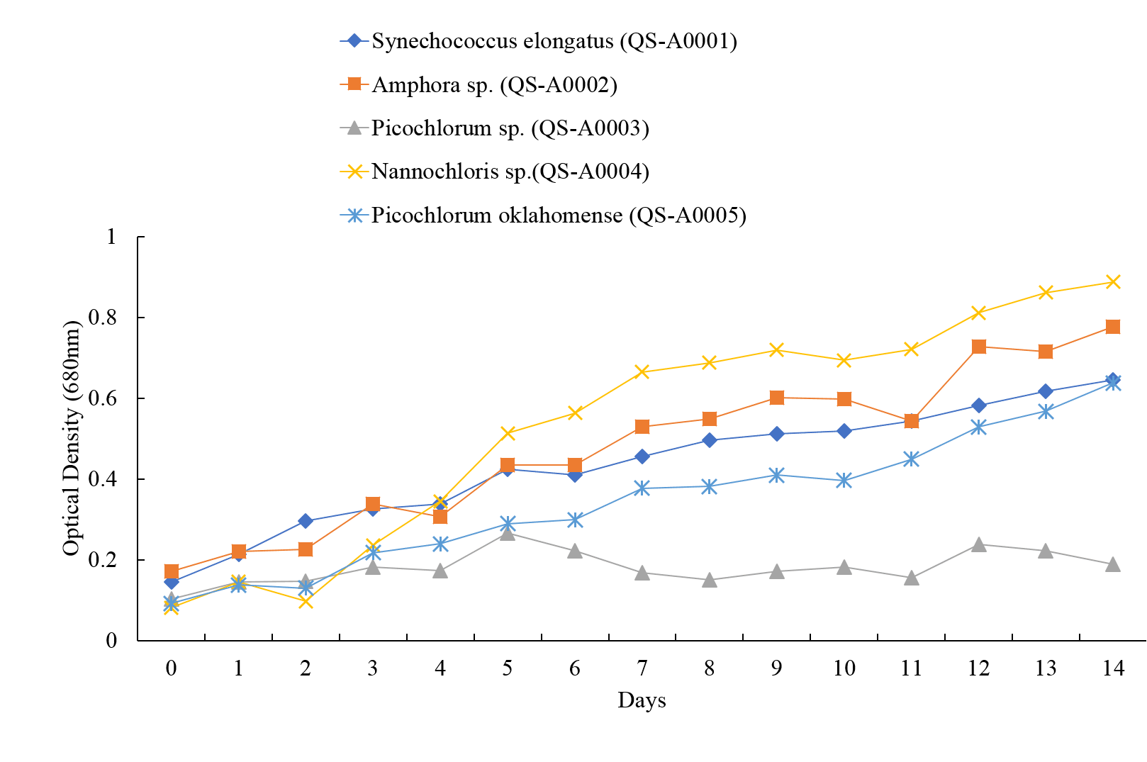
Figure 3. Growth curve of isolated microalgae during 14 days of cultivation.
In this study, measurements taken on day 14 revealed marked differences between planktonic and benthic isolates. While planktonic taxa such as Picochlorum sp., P. oklahomense, Nannochloris sp., and S. elongatus exhibited similar efficiencies, the markedly lower efficiency of Amphora sp. likely reflects methodological bias rather than poor viability (Table 5). Uneven cell distribution and shading effects caused by settled diatoms may mask actual biomass levels (Arnaldo et al., 2024; Prelle et al., 2021; Huang et al., 2019). These limitations highlight the need for alternative or complementary growth assessment methods, such as direct cell counts, in vivo chlorophyll fluorescence, or particulate organic carbon quantification, when evaluating benthic taxa.
On day 14, OD measurements of the five isolated microalgae ranged from 0.20 to 0.67, reflecting differences in adaptation and photosynthetic efficiency. Picochlorum sp. (0.67 ± 0.15), S. elongatus (0.66 ± 0.20), Nannochloris sp. (0.66 ± 0.02), and P. oklahomense (0.61 ± 0.03) showed no significant differences (P > 0.05), indicating comparable growth under the given conditions. Amphora sp., however, recorded significantly lower absorbance (0.20 ± 0.02; P < 0.05). Sedimentation at the bottom of culture vessels and cuvettes reduces light exposure and promotes cell overlap, thereby limiting photosynthetic activity. Consequently, OD-based measurements may underestimate benthic growth, and alternative methods such as direct cell counts or in vivo fluorescence are recommended (Cointet et al., 2019).
Table 5. Optical density of isolated microalgae on day 14 of cultivation.
| Microalgae | Day 14 (OD 680 nm) |
| Synechococcus elongatus | 0.66 ± 0.20a |
| Amphora sp. | 0.20 ± 0.02b |
| Picochlorum sp. | 0.67 ± 0.15a |
| Nannochloris sp. | 0.66 ± 0.02a |
| Picochlorum oklahomense | 0.61 ± 0.03a |
Mean optical density (OD) at 680 nm for five isolated microalgae strains, measured on Day 14 of batch culture, is presented. Values are expressed as mean ± standard deviation (n = 3). Different superscript letters indicate significant differences between groups (P < 0.05).
Beyond methodological considerations, these findings have significant implications for strain selection in aquaculture applications. Planktonic microalgae present operational advantages for large-scale cultivation due to their stable suspension in the water column, a reduced risk of photolimitation, and ease of biomass harvesting. In contrast, benthic species, while potentially valuable for specialized applications such as biofilm-based nutrient removal, may necessitate more complex culture systems to achieve comparable productivity. Therefore, culture design must align with the ecological traits of the target species to maximize yield and functional performance.
4. Conclusions
Five microalgae strains, namely Synechococcus elongatus, Amphora sp., Picochlorum sp., Nannochloris sp., and Picochlorum oklahomense, were isolated from wastewater and pond systems in a commercial shrimp farm. Green microalgae were the most common, found at multiple sites, while cyanobacteria and diatoms were limited to specific locations, likely due to site-specific environmental conditions. All strains showed measurable biomass accumulation over 14 days. Planktonic species maintained higher and more consistent OD values than the benthic diatom Amphora sp., whose lower optical readings were attributed to sedimentation effects rather than poor growth. These differences highlight the influence of morphology on culture performance: planktonic forms are better suited to suspended batch cultures, while benthic species may require modified systems to enhance light access and resuspension. The results indicate that planktonic strains, particularly Picochlorum spp., Nannochloris sp., and S. elongatus, are promising candidates for biomass production and live food applications in aquaculture, while Amphora sp. holds value in benthic or biofilm-based nutrient recovery systems. The diversity of strains recovered demonstrates the potential of aquaculture environments as reservoirs of microalgae with both functional and commercial value.
