


Lumpy skin disease (LSD) is a transboundary animal disease characterized by the formation of skin nodules on the body, along with fever, swollen lymph nodes, reduced milk production, and potential temporary or long-term sterility in bulls (Sprygin et al., 2019). The illness was first documented in Zambia, Africa, in 1929 and remained confined to Africa for many years. However, it made its first transatlantic expansion to Egypt in 1988, followed by an epidemic in Israel in 1989 (Akther et al., 2023). Since then, the disease has spread to parts of Europe, the Middle East, and, more recently, several Asian countries (Pal et al., 2024; Whittle et al., 2023; Ratyotha et al., 2022; Beard et al., 2016).
Poxviruses are double-stranded DNA viruses characterized by terminal hairpin loops at the ends of their genomes. They are identifiable by their distinctive brick-shaped appearance when viewed under an electron microscope (Verma et al., 2024). The lumpy skin virus, part of the Poxviridae family, is a 150 kb encapsulated DNA virus (Kumar et al., 2025). LSDV's genome consists of double-stranded DNA that encodes 30 pox viral proteins, both structural and nonstructural. This virus shares a high degree of genetic and antigenic similarity with sheep pox virus (SPPV) and goat pox virus (GTPV), with nucleotide sequences showing 96% identity across these species (Hamdi et al., 2020).
Bangladesh is an agriculture-based country where rural livelihoods largely depend on livestock rearing, which plays a vital role in socio-economic development (Sobur et al., 2025). The livestock sector contributes approximately 20% of national employment and around 12% of agricultural GDP (Islam et al., 2025; Arefin et al., 2024). The country has an estimated 23.64 million cattle. Given Bangladesh’s geographical proximity to other South Asian countries where LSD is endemic, the disease poses a regional threat to livestock production and food security. In mid-2019, veterinary authorities in Bangladesh identified an outbreak of an unfamiliar syndrome characterized by nodular skin lesions. This outbreak affected both commercial and backyard cattle populations in specific areas of the Chattogram district, including Anwara, Karnaphuli, and Patiya Upazila (Hasib et al., 2021). A similar clinical onset pattern was later observed in various districts throughout the nation (Khalil et al., 2021).
LSD occurs year-round but is more severe in hot, humid climates. The virus is robust and can survive in dried scabs, posing contamination risks. The severity of the disease depends on the virulence of the virus and host factors such as immunity, cattle breed, and age. While cattle are the primary hosts, LSD can also infect buffalo and sheep, although with milder symptoms (Elhaig et al., 2017). Transmission of LSDV through direct contact is inefficient and typically requires parenteral inoculation. Generalized disease may result from arthropod-mediated spread, particularly through intravenous feeding. Insects and ticks, such as Aedes aegypti, Stomoxys calcitrans, and Rhipicephalus appendiculatus, have been identified as effective mechanical vectors for the virus (Paslaru et al., 2021; Rouby et al., 2017; Lubinga et al., 2015).
Clinical signs of LSD include fever, skin nodules, leg swelling, edema, lymph node enlargement, lameness, and cellulitis. Common symptoms also encompass anorexia, reduced milk production, and varying degrees of weight loss. Some animals may experience complications such as mastitis and myiasis (Abutarbush et al., 2015). Bulls can suffer from either permanent or temporary infertility, and the virus can persist in their sperm for extended periods (Sprygin et al., 2019).
The occurrence of LSD varies widely, with reported cases ranging from 2% to 85% in different regions. In endemic areas, the typical morbidity rate is around 100%. LSDV affects cattle of all sexes and ages, but research indicates that young animals are particularly vulnerable to this fatal disease. While the mortality rate is generally low, ranging from 1% to 5%, some reports have indicated higher fatality rates. The morbidity rate in LSD can range from 50% to 100% (Casal et al., 2018).
As a viral disease, LSD lacks a specific treatment. Supportive care may include antibiotics to prevent secondary bacterial infections, anti-inflammatories to reduce fever, and antihistamines to minimize tissue damage. Effective strategies for combating the disease involve widespread cattle vaccination, along with measures such as disinfection, farm biosecurity, stamping-out, and movement controls. However, approaches to eradication and control may differ among countries due to factors like climate and local farming practices. This study aims to estimate the prevalence of LSDV in this Upazila, evaluate associated risk factors, and provide evidence-based recommendations for the treatment, prevention, and control of LSDV.
2. Materials and methods
2.1 Ethical approval statement
No ethical approval was required for this study.
2.2 Study area and periods
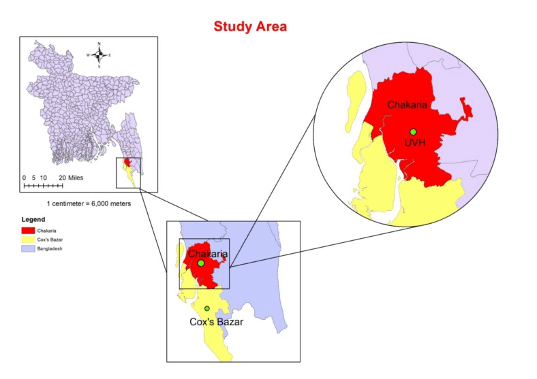
The study was conducted at the Upazila Veterinary Hospital (UVH) in Chakaria, Cox's Bazar, from April to June 2023 (Figure 1).
2.3 Data collection and questionnaire design
A total of 274 clinical cases of cattle were documented during the reporting period at the UVH in Chakaria. Cattle owners initially brought their animals to the hospital and completed a registration process. Subsequently, required information about the patients and owners were collected through a standardized questionnaire. After diagnosing the diseases, appropriate treatments were administered, and regular follow-up assessments were conducted to monitor the progression of the diseases over several days.
2.4 Clinical findings to diagnose LSD
After a visual assessment of the patient, a comprehensive clinical examination was conducted, tailored to the specifics of each case. This examination considered factors such as the patient's disease history, the owner's complaints, and the exhibited symptoms. The diagnosis of LSD cases was based solely on the observed clinical signs and the patient's clinical history. Prominent clinical signs of LSD include high body temperature (105–106 °F), skin nodules, swelling in the legs and brisket, and occasional respiratory distress during critical stages of the disease.
2.5 Inclusion criteria
All cattle presented to the UVH in Chakaria, Cox’s Bazar, during the study period from April to June 2023 were included, regardless of age, sex, or breed, as long as they exhibited clinical manifestations consistent with LSD. The diagnosis was primarily based on characteristic skin nodules identified through close visual inspection and palpation, along with supportive clinical signs such as elevated body temperature (105–106 °F), enlarged superficial lymph nodes, and systemic symptoms including anorexia, reduced milk yield, emaciation, and depression. Additionally, associated complications such as leg and brisket swelling, lameness, respiratory distress, mastitis, and myiasis were considered important diagnostic indicators.
2.6 Exclusion criteria
Cattle were excluded from the study if they exhibited vague or non-specific symptoms that could not be reliably attributed to LSD, or if they had pre-existing dermatological or systemic conditions, such as dermatophilosis, photosensitization, or allergic dermatitis, that could mimic the disease. Additionally, cases with incomplete clinical or epidemiological records, as well as those lacking owner-provided consent or sufficient information in the standardized questionnaire, were also excluded. All animals were examined by trained veterinarians following standardized clinical protocols, and data were systematically recorded through structured questionnaires to ensure the reliability and accuracy of the information collected.
2.7 Data management and statistical analysis
Data was collected from UVH, organized, and stored in a Microsoft Excel 2019 spreadsheet. Descriptive statistics were then utilized for analysis in STATA 16 (Stata Corp, College Station, TX 77845), with p-values ≤ 0.05 considered statistically significant. After assessing the data, I created several tables and figures to illustrate the relationship between disease distribution and factors such as age, sex, breed, herd size, and control measures.
3. Result and Discussion
The study reported a prevalence rate of 62.4%, based on 171 affected cases out of a total of 274 examined animals. The overall mortality rate was 4.34%, with 12 deaths in the total population. Among the affected animals, the case fatality rate was determined to be 7.02%, indicating that 12 of the 171 diseased animals succumbed to their conditions (Figure 2).
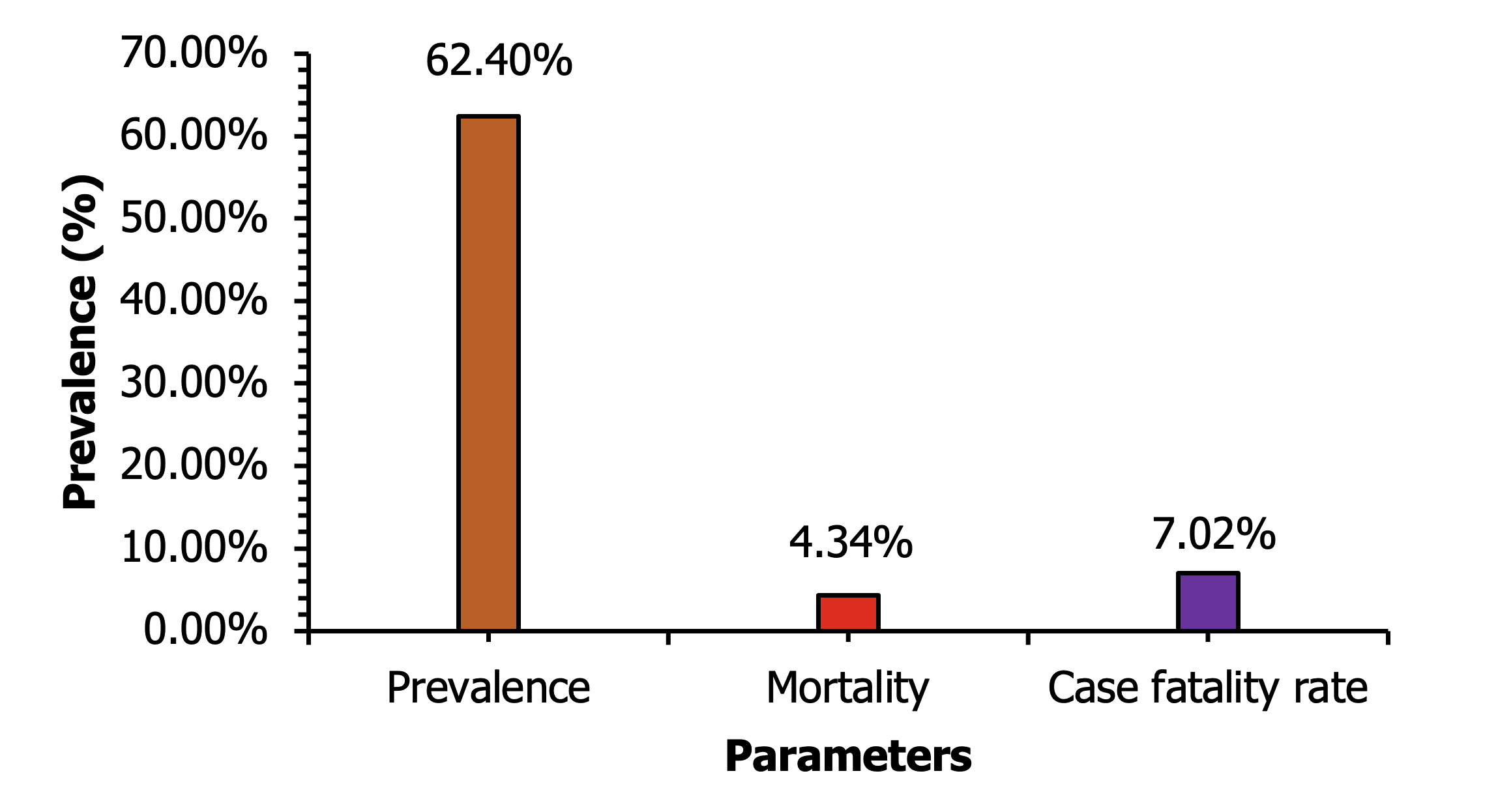
Approximately 62.4% of the cattle in the study were infected with LSD, indicating a relatively high prevalence of the disease among the sampled population. The prevalence of LSD can vary widely, ranging from as low as 6.4% to exceeding 100% (Abebaw et al., 2024). In contrast, Natore Sadar and Baraigram Upazilas recorded significantly higher prevalence rates, affecting approximately 64.70% and 83.02% of cattle, respectively (Haque et al., 2021). A study by Biswas et al. (2020) revealed that Monirampur and Avoynagor Upazilas had rates of 63.33% and 52.38% of affected cattle, respectively. Additionally, another study observed an incidence rate of 49% in their research conducted in Badalgachi, Naogaon (Haque and Gofur, 2020). Another study in Dinajpur, Bangladesh, conducted by Sarkar et al. (2020), reported a prevalence of LSD at 41.06%. Furthermore, Pal et al. (2024) noted that LSD is more prevalent among cattle aged ≤ 24 months in Kaligonj Upazila, Jhenaidah.
The study revealed a 4.34% mortality rate among the studied population, indicating some deaths among the infected animals, which is relevant to other research. The mortality rate was slightly higher during the LSD outbreak in Turkey (Sevik and Dogan, 2017). In Natore Sadar, the mortality rate stood at 2.94%, while in Baraigram, it was slightly higher at 3.77% (Haque et al., 2021). Biswas et al. reported a mortality rate of 1.59% in Abhaynagar and 3.33% in Monirampur Upazila (Biswas et al., 2020). Haque and Gofur found a lower rate of 0.5%, while earlier research indicated mortality rates ranging from 0.99% to 2.12% (Haque and Gofur, 2020).
The survey reported a significant 7.02% case fatality rate, emphasizing the severity of the disease, as 7.02% of infected cattle did not recover. Similar findings were observed in another research. In Natore Sadar and Baraigram, case fatality rates were around 4.53% and 4.55%, respectively (Haque et al., 2021). Biswas et al. indicated rates of 5.26% in Monirampur and 3.03% in Avoynagor, while Haque and Gofur's research reported a lower rate of 1% (Biswas et al., 2020; Haque and Gofur, 2020).
The obtained P value of 0.02 from Table 1 indicates that the chi-squared test signifies a statistically significant link between gender, age group, and animal infection or mortality. This suggests that gender and age are influential factors in determining the likelihood of animals becoming infected and dying due to the infection. The table shows that males have a higher incidence of LSD infection and mortality compared to females. Additionally, the 0-6 months age group demonstrates the highest number of infections and deaths in both male and female animals. Similar results were reported in other studies. Compared to female animals, male animals had a higher rate of infection. This elevated infection rate in males may result from physical fatigue rather than biological factors (Badhy et al., 2021; Abera et al., 2015). Ochwo et al. (2019) also noted higher rates in males and observed sex as significant in Uganda. In contrast, Sarkar et al. (2020), Elhaig et al. (2017), and Molla et al. (2018) found no significant association between sex and LSD.
Table 1. Relation between age and sex of the Lumpy skin disease affected animal.
According to Table 2, the P value is greater than the typical significance level of 0.05, suggesting that there is no significant association between age and limb swelling in cattle infected with LSD. Notably, the 6-12 months age group has the highest percentage of animals with limb swelling (27.78%). Another study found a significant difference in limb swelling among age groups, with the highest incidence (12.02%) observed in young cattle aged 1-3 years (Biswas et al., 2020). Limb swelling and lameness are common clinical manifestations in cattle infected with LSD (Tassew et al., 2018; Gelaye et al., 2015). Lameness is caused by the development of ulcerative skin nodules that extend into tendons and sheaths (Tranquille et al., 2024). Additionally, joint swelling, cellulitis, or phlegmon can lead to arthritis and lameness in cattle (Khan et al., 2024). Nevertheless, some cases of limb swelling may result from age-related factors.
Table 2. Limb swelling according to different age category of infected cattle in the affected area.
According to Table 3, our study results indicate a lower disease risk in herds with indigenous cattle (47.37%) compared to crossbreeds (52.63%). However, another study shows a prevalence of LSD in crossbreeds at 39.2% and in indigenous breeds at 19.2%, possibly due to differing management practices (Begum et al., 2024). While some confounding factors exist, numerous reports support the notion that indigenous cattle have a reduced risk and milder symptoms compared to crossbreds (Saha et al., 2024; Kiplagat et al., 2020). The increased susceptibility of crossbred cattle may be attributed to their lower disease resistance compared to indigenous breeds (Tageldin et al., 2014).
Table 3. Relation between breed and Lumpy skin disease affected animal.
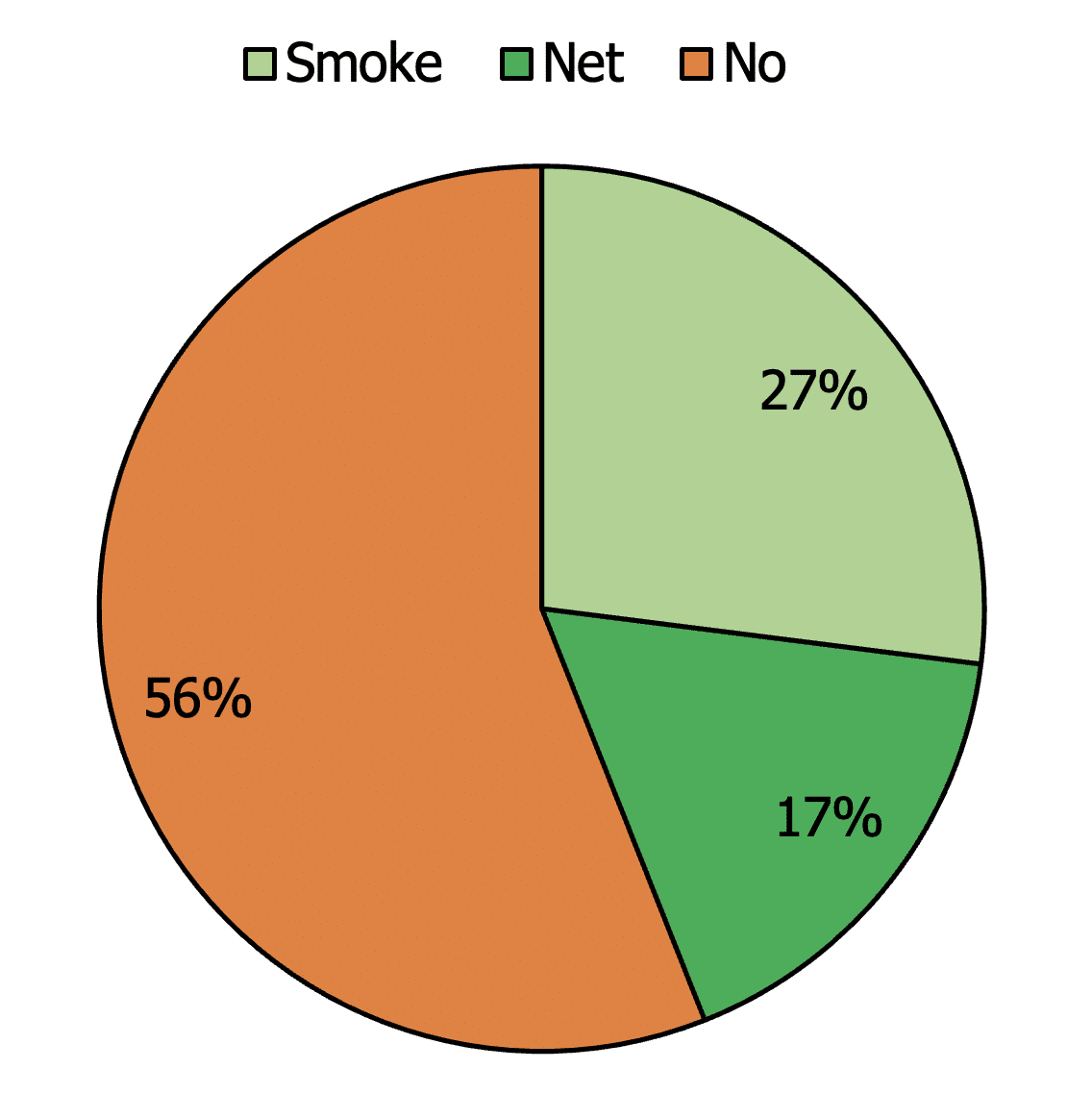
The data illustrates the distribution of vector control methods used to combat the spread of LSD (Figure 3). A notable observation is that a majority (56%) of vector control relies on the "no" control method. This may be attributed to resource constraints, gaps in awareness, or reliance on natural controls. A small group of farmers consciously uses mosquito nets or employs fumes in cowsheds to repel mosquitoes and flies. Other studies have also indicated a lower usage of mosquito nets (Haque et al., 2021; Biswas et al., 2020).
In a study conducted in Chuadanga Sadar, among the examined cattle, 94 (13.62%) showed clinical signs of LSD. Males (17.74%) were more frequently affected than females (10.26%). Crossbred cattle exhibited a higher prevalence (15.22%) compared to indigenous breeds (11.65%). Lesions were most often distributed across the entire body surface (36.17%) (Akter et al., 2025).
Limitations of the study include a short observation period and reliance on hospital-based data, which may not fully represent the entire cattle population in the region. Focusing solely on Chakaria Upazila also restricts the applicability of the findings to other areas. Moreover, the use of hospital-based data may introduce biases and inaccuracies when assessing LSD prevalence and risk factors. Additionally, information reported by farmers may have introduced recall bias, and the limited study period (April–June) may not adequately capture seasonal variations in LSD prevalence.
4. Conclusions
In conclusion, the study found a high prevalence of Lumpy skin disease (LSD) among cattle in the Chakaria Upazila, significantly affecting mortality and case fatality rates. Gender and age were identified as influential factors, with males and younger animals being more susceptible. Indigenous cattle exhibited milder symptoms compared to crossbreeds. The study also revealed limited use of vector control measures, likely due to resource constraints and lack of awareness. Further research and targeted interventions are needed to mitigate the impact of LSD on cattle populations in the region.
Acknowledgements
We express our gratitude to Dr. Mohammad Belayet Hossain for his helpful support during the data collection process. Every farmer's assistance during the research time is much appreciated.
Source of funding
Not applicable.
Data availability
The data generated from this study might be available on the valid request.
Informed consent statement
No informed consent was required to conduct the study.
Conflict of interest
The authors declare no conflict of interest.
Authors’ contribution
Conceptualization, methodology, data collection, and preparing original manuscript: Shiful Islam; Data analysis, reviewing, revising the manuscript: Himangsu Sarker; Data analysis, manuscript formatting: Iqbal Hossain; Data analysis and manuscript formatting: Mohammad Belayet Hossain; Manuscript formatting, reviewing and revising of the final manuscript: Kazi Shams Al Arefin. All authors critically reviewed the manuscript and agreed to submit final version of the article.
